
Overview
The visual and vestibular systems interact to maintain visual clarity of objects during head movement. The reflex that makes this possible is known as the vestibuloocular reflex (VOR) (see the image below). It permits individuals to perform such routine activities as reading street signs while walking down a sidewalk. Like many other systems in the body, most individuals are unaware of the VOR and its basic functioning until it does not function properly. Vertigo, dizziness, imbalance, nausea, vomiting, and other similar symptoms often characterize dysfunction of the vestibular system.
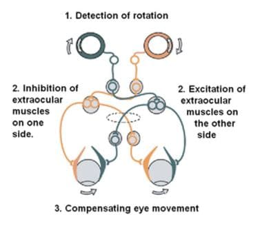
The vestibulo-ocular reflex. A rotation of the head is detected, which triggers an inhibitory signal to the extraocular muscles on one side and an excitatory signal to the muscles on the other side. The result is a compensatory movement of the eyes. Image courtesy of Wikimedia Commons.
Patients who present with symptoms of vestibular dysfunction often undergo a detailed diagnostic workup including a standard electronystagmography (ENG) battery, posturography, and audiometry.
Caloric testing is routinely performed in most vestibular laboratories as part of a vestibular dysfunction workup. Caloric testing is used to assess the integrity of the peripheral vestibular system through stimulation of the horizontal semicircular canals.
The stimulus of warm, cool, or ice water is equivalent to rotational stimulation at a frequency of 0.002-0.004 Hz. These levels of stimulation are significantly lower than those experienced by the VOR system on a daily basis and may not identify dysfunction at higher frequencies.
In addition, caloric results may be abnormal in individuals with congenital abnormalities such as atretic or stenotic external auditory canals, with anatomic variations such as a thickened temporal bone, or with certain acquired disorders such as a severely atelectatic or absent tympanic membrane or fluid in the middle ear. Thus, an additional test is needed to assess the integrity of the VOR.
Rotational chair commonly stimulates frequencies in the 0.01-1.28 Hz range. Head autorotation, an alternative method of testing VOR, stimulates frequencies of 1-6 Hz. This test is discussed in greater detail in the Medscape Reference article Vestibuloocular Reflex Testing.
VOR testing is often performed with the use of a rotary chair. Rotational chair testing was first introduced by Bárány in 1907. He initially designed the chair for VOR testing with impulsive rotation in mind. The test consisted of manual rotation of the chair 10 times over 20 seconds followed by a sudden stop of the chair to analyze the postrotary nystagmus. Rotational chair testing has undergone numerous changes since that time and now has additional applications, including testing of visual-vestibular interaction, optokinetic after-nystagmus (OKAN), high-velocity sinusoidal testing, and off-vertical axis rotation (OVAR). Some of the newer applications require more sophisticated equipment than that developed by Bárány.
Background
As previously stated, the visual and vestibular systems interact to maintain visual clarity of objects during locomotion and other head movement. The fovea (see the image below) is the part of the eye that has the greatest density of photoreceptors and therefore is the area with the best visual acuity. An object is most clearly viewed when it is centered on the fovea. During motion, the image of the viewed object tends to slip from the fovea, causing it to blur. In fact, visual acuity declines to 50% when an object is 2° from the center of the fovea.
To maintain an object on the fovea, the eye must make corrective responses. These corrective eye movements, known as nystagmus, have a slow phase and a quick phase. The vestibuloocular reflex (VOR) slow phase keeps the eyes on the foveal vision (opposite direction to head movement).
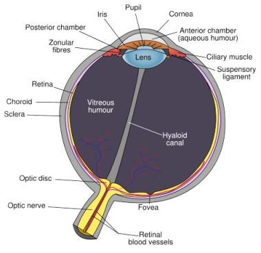
Schematic diagram of the human eye, with the fovea at the bottom. Image courtesy of Wikimedia Commons.
Many different types of nystagmus exist, some of which are physiologic and others of which are pathologic. During rotational chair testing, the alert patient’s eyes move in a direction opposite to the rotation of the chair. As the globe reaches an eccentric position within the orbit, a corrective response attempts to move it back to the center of the orbit. The nystagmus that is observed is a physiologic response and is observed with acceleration and deceleration of rotation. Sustained rotation results in a decline of this nystagmus. Multiple methods of rotation exist, each of which was designed to analyze vestibular responses by observation of the eye movements.
The rotational chair has primarily been used for analyzing horizontal canal VOR.
Rotation of the chair is performed with the assumption that the stimulus applied to the whole body is the same as a stimulus that is applied to the head. Therefore, the head should be secured to the chair during rotation. In addition, most commercially available test protocols do not exceed a frequency of 1 Hz because the skin may move relative to the skull at frequencies greater than 1 Hz, thereby nullifying the assumption.
Relevant Anatomy
The vestibular system, which is the system of balance, consists of 5 distinct end organs: 3 semicircular canals that are sensitive to angular accelerations (head rotations) and 2 otolith organs that are sensitive to linear (or straight-line) accelerations.
The semicircular canals (see the image below) are arranged as a set of 3 mutually orthogonal sensors; that is, each canal is at a right angle to the other 2. This is similar to the way 3 sides of a box meet at each corner and are at a right angle to one another. Furthermore, each canal is maximally sensitive to rotations that lie in the plane of the canal. The result of this arrangement is that 3 canals can uniquely specify the direction and amplitude of any arbitrary head rotation. The canals are organized into functional pairs wherein both members of the pair lie in the same plane. Any rotation in that plane is excitatory to one of the members of the pair and inhibitory to the other.
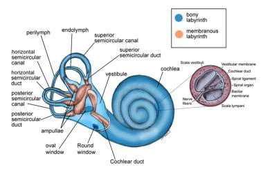
Anatomy of the labyrinth.
For more information about the relevant anatomy, see Vestibular System Anatomy, Visual System Anatomy, Extraocular Muscle Actions, Extraocular Muscle Anatomy, and Inner Ear Anatomy. Also see Vestibuloocular Reflex Testing.