
Practice Essentials
Osteogenesis imperfecta (OI) is a disorder of bone fragility chiefly caused by mutations in the COL1A1 and COL1A2 genes that encode type I procollagen.
Four types of osteogenesis imperfecta were originally described by Sillence in 1979 and are now used broadly as the Sillence Criteria.
The Nosology and Classification of Genetic Skeletal Disorders provides similar categorization in the 2010 revision.
Precise typing is often difficult, and depends in large degree on the experience of the clinician. Severity ranges from mild forms to lethal forms in the perinatal period. Additional genes have been discovered in which mutations can also cause brittle bones. These are typically clinically indistinguishable and are considered by most to be subtypes of osteogenesis imperfecta.
Examples of common radiologic findings of osteogenesis imperfecta are shown in the images below.
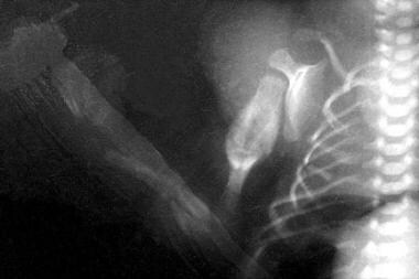
Acute fractures are observed in the radius and ulna. Multiple fractures can be seen in the ribs. Old healing humeral fracture with callus formation is observed.
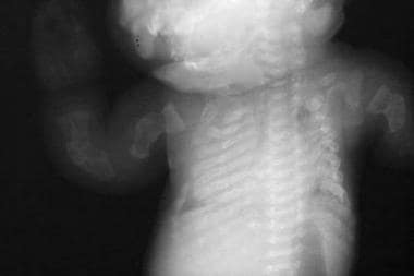
Beaded ribs. Multiple fractures are seen in the long bones of the upper extremities.
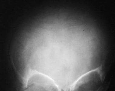
Wormian bones are present in the skull.
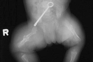
This newborn has bilateral femoral fractures.
Physical characteristics of osteogenesis imperfecta
Individuals with type III (severe, progressively deforming) OI may have joint hyperlaxity; muscle weakness; chronic, unremitting bone pain; and skull deformities (eg, posterior flattening) due to bone fragility during infancy. Limb shortening and progressive deformities can occur, and patients have a triangular face with frontal and temporal bossing; malocclusion is common. The sclera have variable hues.
Workup in osteogenesis imperfecta
Results from routine laboratory studies in patients with OI are usually within reference ranges, and they are useful in ruling out other metabolic bone diseases, such as hypophosphatasia or inherited forms of rickets.
Collagen synthesis analysis has historically been performed by culturing dermal fibroblasts obtained during skin biopsy. This testing modality has fallen out of favor, given that molecular diagnostic techniques are more accurate, have increased in availability, and have decreased in cost. Widespread adoption of next-generation sequencing (NGS) technology has allowed for parallel sequencing of many genes. Thus, there is little or no difference in cost or turnaround time for multigene panels compared with limiting investigation to the two procollagen I genes.
Obtain a radiographic skeletal survey after birth. Prenatal ultrasonography can be used to detect limb-length abnormalities at 15-18 weeks’ gestation.
The width of biopsy cores, the width of the cortex, and the volume of cancellous bone are decreased in all types of osteogenesis imperfecta. The number and thickness of trabeculae are reduced.
Management
Cyclic administration of intravenous pamidronate reduces the incidence of fracture and increases bone mineral density, while reducing pain and increasing energy levels.
Intravenous zoledronate has been increasingly used in children with OI, given its shorter infusion times and longer infusion intervals. Observational studies have shown similar effects to that of pamidronate, with increased bone mineral density, decreased fracture rate, and improvement in pain.
There is scant evidence supporting the use of bisphosphonates in adults with OI, although they continue to be widely used; anecdotally, some patients experience a subjective decrease in pain.
The bisphosphonate risedronate may have some effect in reducing fractures in patients with OI.
Nutritional evaluation and intervention are paramount to ensure appropriate intake of calcium and vitamin D. Caloric management is important, particularly in adolescents and adults with severe forms of OI.
Orthopedic surgery is one of the pillars of treatment for patients with OI.
Surgical interventions include intramedullary rod placement, surgery to manage basilar impression, and correction of scoliosis.