
Overview
Dysplastic (atypical, Clark) melanocytic nevi are acquired pigmented melanocytic proliferations of the skin with distinct clinical and histologic features. In the appropriate clinically setting dysplastic (atypical, Clark) melanocytic nevi are cutaneous markers for CDKN2A mutation and the development of familial and nonfamilial melanomas.
Dysplastic melanocytic nevi, Clark nevi, and nevus with architectural disorder (NAD) are synonymous terms to describe this acquired melanocytic proliferation. The National Institutes of Health (NIH) consensus conference recommended these lesions be defined clinically as atypical nevi and histologically as NAD (see the following image), as well as assigned a grade of melanocyte atypia as mild, moderate, and severe, although some authorities prefer to divide as high or low grade, or do not grade at all.
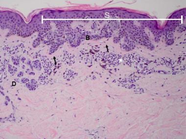
Pathology of dysplastic (atypical, Clark) melanocytic nevi. Compound melanocytic nevus with architectural disorder and shouldering (S), or extension of the junctional component beyond the dermal nests of melanocytes (D). Rete ridges are irregular and distorted with bridging (B) and eosinophilic fibrosis (arrows). Scattered lymphocytic infiltrate is often present (*).
Dysplastic (atypical, Clark) melanocytic nevi appear most commonly on the trunk. Similar lesions on acral sites, breast, genitalia, and the scalp and forehead in children are not associated with the familial syndrome but share histological features with Clark nevi.
See Atypical Mole (Dysplastic Nevus) for more information.