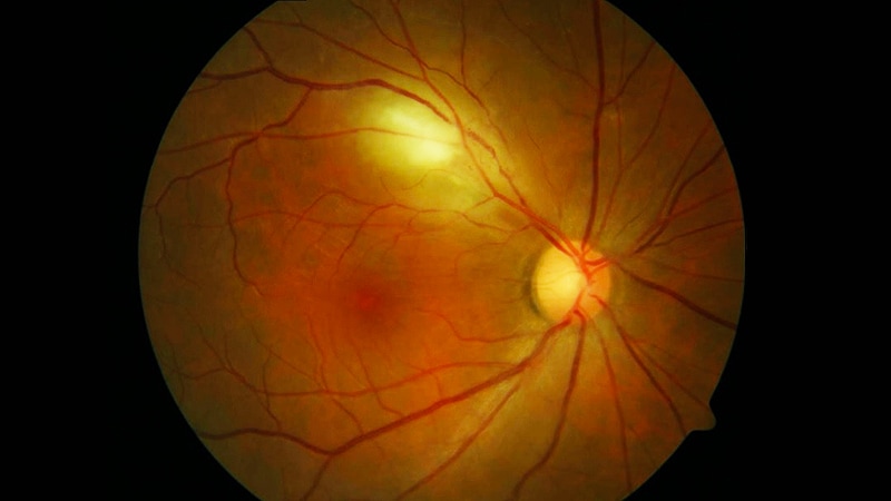
An eye exam may be all that is needed to diagnose Parkinson’s disease, new research shows.

Maximillian Diaz
Using an advanced machine-learning algorithm and fundus eye images, which depict the small blood vessels and more at the back of the eye, investigators are able to classify patients with Parkinson’s disease compared against a control group. “We discovered that micro blood vessels decreased in both size and number in patients with Parkinson’s,” Maximillian Diaz, a PhD student at the University of Florida in Gainesville, told Medscape.
The simple eye examination may offer a way to diagnose Parkinson’s early in the disease progression.
Diaz said the test could be incorporated to a patient’s annual physical examination not only to look for Parkinson’s but also for other neurological diseases. A team in his lab is also looking at whether the same technique can diagnose Alzheimer’s disease.
The beauty of this is that “the technique is simple,” he said. “What surprised us is that we can do this with fundus images, which can be taken in a clinical setting with a lens that attaches to your smartphone.”
“It’s affordable and portable and it takes less than a minute,” he added.
Machine Learning on Fundus Eye Images
Researchers, under the direction of Ruogu Fang, PhD, director of the J. Crayton Pruitt Department of Biomedical Engineering’s Smart Medical Informatics Learning and Evaluation Lab (SMILE), collected fundus eye images from 476 age- and gender-matched individuals, 238 diagnosed with Parkinson’s and 238 control group images. Another set of 100 images were collected from the University of Florida database using green color channels (UKB-Green and UF-UKB Green) and used to improve vessel segmentation. Of these, 28 were controls and 72 from Parkinson’s patients. Researchers added 44 more control images from the UK Biobank to complete the second age- and gender-matched dataset.
“We used 80% of the images to develop the machine-learning network,” Diaz said. The other 20% of images, which were new to the algorithm, were used to test it, to determine true or false, Parkinson’s or control?
“We were able to achieve an accuracy of 85%,” Diaz told Medscape Medical News. Currently, there are no biomarkers to diagnose Parkinson’s. The disease is only recognizable once 80% of dopaminergic cells have already decayed. “Clinically, there’s no way to tell how long someone has had it,” Diaz said. He hopes that by doing additional research and testing earlier — with a longitudinal study of images — a pattern may be detected to better predict disease.
Eye Vasculature Reveals Disease
“This concept [studying eye vasculature] is getting a lot of interest right now,” Anant Madabhushi, PhD, Case Western Reserve University, Cleveland, Ohio, told Medscape Medical News. “The eye is the proverbial window to the soul, and in this case, shows what’s happening in rest of the body.”
Madabhushi, who was not involved in the Parkinson’s research, has also been working with a team in Cleveland to look at how vessels in the eye predict response to drug therapies in diabetic macular edema, including treatment durability. “What we’ve found is the more twisted the vessels, the more constricted, and the less likely the person would respond to therapy,” he said, adding that studying the pathology of the eye makes a lot of sense. “The arrangement of vessels in the eye are likely to have implications in all kinds of diseases.”
Since Parkinson’s disease does not have any biomarkers, this technology could be very useful in diagnosis. “With specific quantitative measurements, we could have computational imaging biomarkers to predict the risk of onset of Parkinson’s, and the prognosis of disease. That’s the true utility of this approach,” he said.
Diaz has disclosed no relevant financial relationships. Madabhushi has consulted for Aiforia Inc and has had research sponsored by AstraZeneca, Bristol-Myers Squibb, and Boehringer Ingleheim.
Radiological Society of North America (RSNA) Annual Meeting: Poster IN-1A-07. Presented November 29, 2020.
Ingrid Hein is a freelance health and technology reporter based in Hudson, Quebec.

