
Osteology
The orbit, which protects, supports, and maximizes the function of the eye, is shaped like a quadrilateral pyramid, with its base in plane with the orbital rim. Seven bones conjoin to form the orbital structure, as shown in the image below.
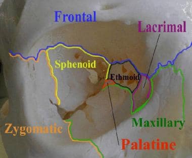
This image of the right orbit shows the 7 bones that contribute to its structure.
The orbital process of the frontal bone and the lesser wing of the sphenoid form the orbital roof. The orbital plate of the maxilla joins the orbital plate of the zygoma and the orbital plate of the palatine bones to form the floor. Medially, the orbital wall consists of the frontal process of the maxilla, the lacrimal bone, the sphenoid, and the thin lamina papyracea of the ethmoid. The lateral wall is formed by the lesser and greater wings of the sphenoid and the zygoma.
The orbits are aligned so that the medial walls are parallel and the lateral walls are perpendicular.
The arc from medial to lateral wall in each orbit is 45°. Lines dropped through a central anterior-to-posterior axis of each orbit bisect at a 45° angle. The floor is two-thirds the depth of the orbit. The average dimensions of the orbit are as follows:
Height of orbital margin – 40 mm
Width of orbital margin – 35 mm
Depth of orbit – 40-50 mm
Interorbital distance – 25 mm
Volume of orbit – 30 cm3
The superficial bony orbit is defined by the orbital margin, which is rectangular with rounded corners. The margin is discontinuous at the lacrimal fossa. The supraorbital notch (seen in the image below) is within the supraorbital rim and is closed to form the supraorbital foramen in 25% of individuals. The supratrochlear notch is medial to the supraorbital notch.
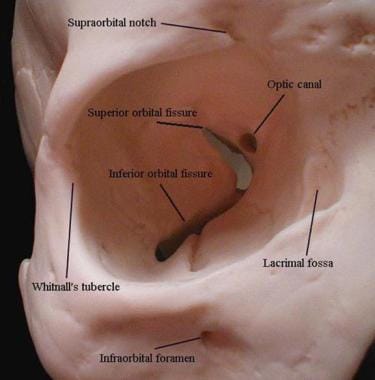
This image of the right orbit shows superficial landmarks, optic canal, and superior and inferior orbital fissures.
The trochlea is a cartilaginous ring that supports the superior oblique muscle. The trochlea attaches to the periorbita within the fovea trochlearis along the superior-medial orbit. The infraorbital foramen is located 10 mm inferior to the zygomaxillary suture.
Laterally, the orbital rim is marked by the Whitnall tubercle, which is 10 mm inferior to the zygomaticofrontal suture. The tubercle is the site of attachment of the lateral canthal tendon.
Orbital fissures and optic canal
The major nerves and vessels to the orbit and globe enter through 3 openings. The superior orbital fissure is bounded by the lesser and greater wings of the sphenoid. The greater wing of the sphenoid, the maxilla, and the palatine bones of the orbit form the boundaries of the inferior orbital fissure. The optic canal is at the apex of the orbit and lies within the sphenoid bone.
The structures entering through the superior orbital fissure are as follows:
Cranial nerves (CN) III, IV, and VI
Lacrimal nerve
Frontal nerve
Nasociliary nerve
Orbital branch of middle meningeal artery
Recurrent branch of lacrimal artery
Superior orbital vein
Superior ophthalmic vein
The structures entering through the inferior orbital fissure are as follows:
Zygomatic nerve
Parasympathetics to lacrimal gland
Infraorbital artery
Infraorbital vein
Inferior ophthalmic vein branch to pterygoid plexus
The structures entering through the optic canal are as follows:
Optic nerve
Ophthalmic artery
Central retinal vein
Ethmoid arteries
The anterior and posterior ethmoid foramina lie in the medial wall of the orbit along the frontoethmoidal suture line. The anterior and posterior ethmoid arteries pass through these foramina and are important surgical landmarks. The arteries mark the level of the cribriform plate and the relationship of the anterior cranial fossa to the orbits. The ethmoid arteries mark the superior limit for osteotomies during medial maxillectomy.
The distance from the orbital rim to the anterior ethmoid artery is approximately 20-25 mm. The distance between the anterior and posterior ethmoid arteries averages 12 mm, with a range of 8-19 mm. The optic ring averages 6 mm from the posterior ethmoid artery, with a range of 5-11 mm. Knowledge of these distances safely guides the surgeon along the medial orbital wall.
Infraorbital foramen
The infraorbital sulcus crosses the floor of the orbit and carries the infraorbital artery, infraorbital vein, and infraorbital nerve from the inferior orbital fissure to the infraorbital foramen. Clinically, the infraorbital foramen provides a route of spread for infection or maxillary tumors to the orbit and the skull base. The surgeon must avoid injury to the infraorbital neurovascular bundle when performing a midface degloving approach.