
Background
Approximately 20% of patients with severe facial trauma injuries also have an ophthalmic injury. In such patients in the ED , eye injuries should be quickly ruled out, including retrobulbar hemorrhage and penetrating globe injury.
Blow-out fractures occur when a blow to the eye increases pressure in the orbit, causing the weak floor or medial wall (lamina papyracea) to “blow out” into the maxillary sinus or ethmoid bone.
This results in a fracture, though it often prevents globe rupture and loss of the eye.
Periorbital fat and extraocular muscles can become entrapped in the fracture, leading to problems of ocular movement.
When the medial wall (lamina papyracea) is fractured, the medial rectus becomes entrapped, leading to lateral gaze dysfunction.
In maxillary fracture, the orbit floor blows out, and the inferior rectus entrapment leads to problems in upward gaze.
The eye can be injured during compression before the ethmoid bone or the maxillary sinus fractures. About one third of blow-out fractures have an associated eye injury.
Superior orbital rim fracture is a frontal bone fracture that is associated with high-impact injuries to the brain, face, and cervical spine.
Tripod fractures and zygomaticomaxillary complex fractures occur from high-impact injury to the cheek’s malar eminence (zygoma). Often, these fractures are associated with eye and inferior orbital nerve injuries.
The orbit is composed of 7 facial bones: frontal, zygoma, maxilla, lacrimal, ethmoid, sphenoid and palatine. The superior orbital ridge and upper medial orbital ridge are part of the frontal bone. The lateral orbital rim is part of the zygoma. The inferior and lower medial rims are part of the maxilla, and the floor of the orbit is made of the upper border of the maxillary sinus.
The medial rim separating the orbit from nares is the lacrimal bone. The medial wall and part of the posterior wall of the orbit are formed by the ethmoid bone. The remainder of the posterior of the orbit is formed by the 2 wings of the sphenoid bone, the continuation of the lacrimal bone from the medial wall, and the orbital process of the palatine bone.
The optic nerve exits the optic foramen in the lesser wing of the sphenoid bone. The globe of the eye sits within the orbit surrounded by periorbital fat and the extraocular muscles that control its movement. The inferior orbital nerve courses through the maxilla in the orbital floor. The weakest portion of the orbit is the thin orbital floor (maxilla) and the lamina papyracea (ethmoid bone) medially and inferiorly.
A study by Coon et al, identified 4 indications for surgical intervention in pediatric patients with orbital fractures
:
Rectus muscle entrapment
Early enophthalmos
Central-gaze diplopia or extraocular movement restriction after the resolution of swelling
Loss of orbital support likely to produce secondary changes in globe position and/or binocular stereo vision
See the image below.
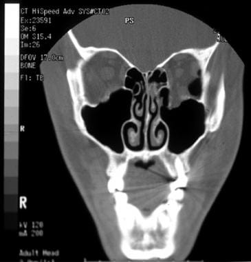
Left orbital floor fracture. This patient presented with little motility disturbance; however, because of the large defect in the orbital floor, late enophthalmos was predicted. Surgical repair was undertaken. Note the pneumo-orbitum.