
Practice Essentials
Rheumatoid arthritis (RA) is a chronic multisystemic disease of unknown cause. The characteristic feature is persistent inflammatory synovitis usually involving peripheral joints in a symmetrical distribution. Synovial inflammation causes cartilage destruction and bone erosion. Subsequently, joint deformity occurs. The axial skeleton, with the exception of the cervical spine, is affected later and less frequently. The risk factors for cervical spine involvement include frequency of peripheral joint erosion, rheumatoid factor (RF) positivity, use of corticosteroids, long RA duration, and markers of higher disease activity (ESR, CRP, and Disease Activity Score).
Clinically, cervical spine involvement in RA patients is reported to be mainly seen in 3 forms: atlantoaxial subluxation (AAS) (65%), superior odontoid migration (SOM) (20%), and subaxial subluxation (SAS) (15%).
In cases of AAS, anterior subluxation may be most common (70%), followed by lateral subluxation (20%). Posterior subluxation is seen in 7% of cases, and rotational subluxation is very rarely reported.
Preferred examination
The mainstay of imaging the rheumatoid spine remains plain radiography to identify anterior atlantoaxial subluxation via the anterior or posterior atlantodental interval; vertical subluxation via the McGregor method or Ranawat index; diameter of the spinal canal; and cervical height index. Flexion/extension views are necessary to assess the level of involvement and any evidence of instability. The need for further imaging by means of CT, MRI, or myelography may also be assessed during radiography.
The major role for CT and MRI is in the preoperative assessment.
The correlation of MRI findings with symptoms is crucial before any surgical decisions can be made. There is a close correlation between the severity of cervical myelopathy and the degree of compression as demonstrated by MRI. Besides influencing the selection of patients for spinal surgery, MRIs can help in planning surgical procedures, especially those in patients with more than 2 levels of cord compression.
Computed tomography allows for a very precise assessment of bone structure of the whole cervical spine. The anatomy of the atlantoaxial joint is more visible than on plain radiograph, where the image deteriorates because of overlapping bone structures. CT is also considered to be the best method for the visualization of bone erosion, as well as atypical subluxation within the atlantoaxial joint, in particular lateral and rotary subluxation.
(See the images below.)
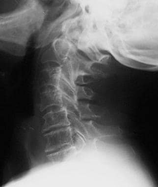
Lateral view of the cervical spine in a patient with rheumatoid arthritis shows erosion of the odontoid process.
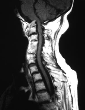
T1-weighted sagittal MRI of the cervical spine shows basilar invagination with cranial migration of an eroded odontoid peg. There is minimal pannus. The tip of the peg indents the medulla, and there is narrowing of the foramen magnum due to the presence of the peg. Inflammatory fusion of several cervical vertebral bodies is shown.
After the metacarpophalangeal joints, the most common region to be involved in RA is the cervical spine. This can lead to severe pain and disability, as well as various neurologic manifestations, although some patients with significant radiographic evidence of disease may be entirely asymptomatic. The frequency of radiographic signs of involvement of the cervical area is 44-86%, depending on the duration of the disease.
RA activity in the cervical spine begins early, with 83% of patients in prospective studies developing anterior atlantoaxial subluxation within 2 years of disease onset. Activity in the cervical spine progresses clinically and radiologically in tandem with peripheral joint involvement. In fact, the severity of the peripheral erosive damage is strongly correlated with the degree of structural damage in the cervical spine. Features of spinal involvement in RA include erosive synovitis, ligamentous subluxation, osteopenia, and vertebral-body fracture.
See Rheumatoid Arthritis: In and Out of the Joint, a Critical Images slideshow, to help identify the distinguishing features of RA as well as the signs of extra-articular manifestations of this disfiguring disease.