
Practice Essentials
Although nasal fractures are the most common facial fracture in both adults and children,
they often go unnoticed by physicians and patients. Patients with nasal fractures usually present with some combination of deformity, tenderness, hemorrhage, edema, ecchymosis, instability, and crepitation; however, these features may not be present or may be transient.
To further complicate the matter, edema can mask underlying nasal deformity, crepitation, and instability; thus, many physicians and patients fail to pursue further diagnosis and appropriate treatment.
Radiologic features of nasal fractures are demonstrated below.
If radiographic evaluation is warranted, it is best used when other facial fractures are suspected in combination with a nasal fracture, because isolated nasal fractures are treated on the basis of the physical examination alone. The fact that patients may have displaced nasal fractures and normal-appearing plain radiographic findings should be emphasized.
Preferred examination
Although the use of plain images is not suggested, the preferred examination includes the acquisition of Waters (occipitomental) and lateral nasal views if plain films are used. It should be noted that plain radiographs only serve to confuse the clinical picture in most cases. Plain radiographs do not allow identification of cartilaginous disruptions, fractures, shearing, and injury in general. Plain radiographs also do not provide sufficient information to assess injury severity and displacement, 2 important aspects essential to emergent and delayed management and surgical planning.
A good physical examination of the internal and external nose is still the method of choice for detecting and assessing nasal fractures.
For questionable fractures and fractures that may be associated with other facial trauma, a CT scan of the facial bones without contrast is an excellent choice. Modern multidetector CT scanners have revolutionized trauma imaging and provide a fast, safe, cost-effective, and sensitive means for assessing trauma of the bone and soft tissues. Three-dimensional reconstructions are easily derived from these scanners and readily show the degree of displacement if substantial trauma exists.
Ultrasonography may be able to detect local and superficial fractures, but it can be difficult to see the entire nasal bane and neighboring bones with ultrasound.
A definitive diagnosis of nasal fracture (nasal dorsum and nasal wall) may be made, when necessary, on the basis of all clinical data combined with x-ray findings (nasal bones and Waters view) and CT scans.
However, x-rays only have an 82% reliability rate.
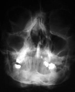
Nasal fractures. Posteroanterior view shows displaced septum from the maxillary crest and a deviated nasal root to the patient’s right.
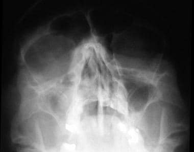
Nasal fractures. Waters view shows a deviated nasal septum, quadrangular cartilage displaced from the maxillary crest, and a nasal root deviated to the right.
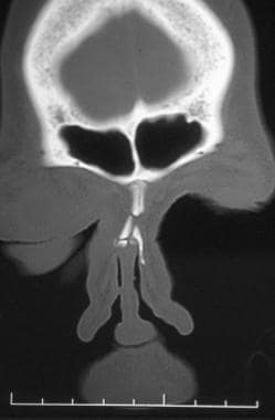
Nasal fractures. Coronal CT scan demonstrates a nasal fracture with root deviation to the right. Note that the fracture has occurred in the weaker lower portion of the nasal bones.
If untreated, nasal fractures can result in unfavorable appearance and function, especially when the underlying structural integrity of bone and cartilage is lost.
Untreated nasal fractures account for the high percentage of rhinoplasty and septoplasty procedures performed months to years after the initial trauma occurs. Thus, appropriate treatment is best rendered in a timely manner, before scarring and soft tissue changes occur. As always, thorough history taking and physical examination should precede radiographic evaluation.
Pathophysiology
Nasal bone fractures account for 50% of all facial fractures, with the majority of these injuries involving the cartilaginous lower two thirds of the nose.
Lateral impact injuries are the most common type of nasal injury leading to fracture.
Nasal fracture and displacement without septal fracture usually occur with weaker applied forces; however, with increased force, displacement of the bilateral nasal bones may be noted, and the septum is usually dislocated and fractured as well.
Approximately 80% of fractures occur at the lower one third to one half of the nasal bones. This area represents a transition zone between the thicker proximal and thinner distal segments.
Other injuries that are commonly associated with nasal fractures include midface injuries involving the frontal, ethmoid, and lacrimal bones; nasoorbital ethmoid fractures; orbital wall fractures; cribriform plate fractures; frontal sinus fractures
; and maxillary Le Fort I, II, and III fractures.
Epidemiology
In the US, approximately 50,000 people experience nasal fractures each year.
Nasal fractures account for approximately 40% of all bone injuries. Fights and sports injuries are the most common causes of nasal fractures in adults, followed by falls and vehicle crashes. Play and sports account for the majority of nasal fractures in children. Physical abuse should be considered when evaluating children and women with nasal fractures. Nasal fractures may occur in isolation, but are commonly associated with other facial injuries and fractures.
Anatomy
The nose is the most prominent and anterior facial feature; as such, it is also the most readily exposed to trauma. The nose is supported by cartilage on the dorsum and caudal aspects and by bone posteriorly and superiorly. The paired nasal bones, the maxillary crest, and the nasal bones, jutting from the frontal bone, form the bony framework that supports the underlying nasal cartilages. The entire lower two thirds of the nose is cartilaginous.
Overlying the framework of the nose are soft tissues, including muscles, nerves, and mucous glands. The lower two thirds of the nose houses 2 upper lateral cartilages, which originate from underneath the inferior aspect of the nasal bones and project into the scroll region of the nose just superior to the nasal tip. The paired upper lateral cartilages are continuous with the dorsal nasal septum. The septum has a dorsal, caudal, posterior, and maxillary attachment as its components and is often referred to as the quadrangular cartilage. The lower lateral crura are composed of the medial, intermediate, and lateral crural components. The medial crura attach to the septum with adhesive ligaments at the caudal septum, giving structural support to the nasal tip, which is composed of the transition between the intermediate and lateral crura.
Limitations of techniques
The use of plain images and computed tomography (CT) scans for the diagnosis and management of nasal fractures has been controversial. Several small studies have shown that use of these modalities is neither cost-effective nor beneficial to the patient or physician. Nasal fractures are usually evident and can be elicited by means of careful history taking and physical examination. Rarely is the radiologic confirmation of these injuries needed.
However, some clinicians still use plain images and CT scans, and the radiologist must understand some of the diagnostic pitfalls to reduce the rate of erroneous readings.
Wexler opined that nasal radiographs were a medicolegal necessity and considered them an important adjunct in acute injuries to the nasal bones.
Unfortunately, this opinion was not founded on solid experimental data. The practice of ordering unnecessary radiographs encourages poor patient care and devalues the importance of thorough history taking and physical examination. This practice also leads to needless irradiation, expense, and wasted time.
Lee et al compared high-resolution ultrasonography (HRUS) with CT in diagnosing nasal fractures in 140 patients with nasal trauma from 2004-2007. The accuracy rates for HRUS, CT, and conventional radiography were 100%, 92.1%, and 78.6%, respectively. Compared with HRUS, CT revealed only 196 of 233 lateral nasal bone fractures. In high-grade fractures, the accuracy of CT was 87%, but it decreased to 68% in low-grade fractures.
De Lacey et al concluded in a study that a lateral plain radiographic view was unreliable for the evaluation of nasal fractures because of the high incidence of similar defects found in noses from control subjects and in patients with dry skulls, when evaluated using plain radiography. In their study, the investigators evaluated 100 consecutive patients presenting to the emergency department with a history of trauma to the nose.
Nasal radiographs were obtained in each patient, including the Waters and lateral views. There were no radiographic findings of fracture in 65 of the 100 patients. Nasal fractures were depicted in 45 patients, yet only 3 patients required reduction, and 31 of 45 patients were discharged without treatment. The authors then compared the lateral radiographs in 50 control subjects and 50 persons with dry skulls.
When images from control subjects were compared with images of persons with dry skulls, misreads were identified and classified as midline defects, high lateral-wall defects, and low lateral-wall defects. In 50 control subjects, 33 cortical defects were observed. After close inspection, the misreads were found to be the result of the midline nasal suture, the nasomaxillary suture (low defect), and thinning of the nasal wall (high defect).
Clayton and Lesser concluded that examination under anesthesia provided more accurate information about nasal injury than did radiography or clinical examination alone or together. The authors prospectively evaluated 54 patients clinically, radiologically, and under anesthesia within 19 days after nasal injury.
Waters views and lateral views were obtained in all patients. External examination and nasal rhinoscopy were performed to evaluate the patients clinically.
The authors not only concluded that examination under anesthesia provided more accurate information than did the other examination methods, they also found that the Waters view, the lateral view, or the 2 views in combination were less useful than was physical examination alone. Standard radiographs were not helpful in deciding whether to perform manipulation for repair or when and how such manipulation should be performed.
When evaluation of children is necessary and one wishes to limit exposure to radiation, ultrasonography has been helpful to some in evaluating nasal fractures, septal deviation, and level of comminution. This can be accomplished with a 7-15 MHz linear array transducer.
In the utilization of 3-dimensional (3D) CT scanning for facial and nasal fractures, better evaluation scores were achieved with surface rendering protocols than with volume rendering protocols. Surface rendering offered better overall image quality than did volume rendering.
The legal value of an examination depends on the degree of medical findings supported by the examination results.
In isolated cases of nasal trauma, radiographs have a high number of false-negative results and a large, but unknown, number of false-positive results. Thus, the legal value is low because of the uncertain degree of confidence in the findings. Radiographic examinations of the nose have been known to fail in the assessment of nasal fractures.