
Practice Essentials
Thyroid nodules are common, perhaps existing in almost half the population, as determined using ultrasonography (US). Only 4-7% of thyroid nodules detected with US are palpable in the adult population in the United States, with women affected more frequently than men. Although the thyroid is the most common endocrine organ to undergo malignant degeneration, thyroid carcinoma accounts for only 1% of diagnosed neoplasms in the United States each year.
Thyroid cancer is rare; the annual detection rate of clinically significant thyroid cancer in the general population is only 0.004%. Only 5-10% of thyroid cancers are clinically palpable.
In a study by Davies et al utilizing data from the National Cancer Institute’s Surveillance, Epidemiology, and End Results Program and the National Vital Statistics System, the rate of thyroid cancer in US adults was found to have risen from 4.9 per 100,000 individuals to 14.3 per 100,000 between 1975 and 2009, and in women, the increase in thyroid cancer was almost 4 times that in men.
Palpable nodules can be visualized as areas of increased (hot) or decreased (cold) tracer activity. However, terms such as hot nodules and cold nodules are misleading unless the nodule can be clearly delineated in several projections. A nonfunctioning (cold) nodule placed in the center of a lobe with functioning normal tissue superficial to it may appear as warm on scans because of integrated activity with depth. Unless oblique views are imaged, the presence of activity concentration cannot be definitively determined. Therefore, nodules should be identified as being functioning (hot), nonfunctioning (cold), or photon deficient. Hot nodules (which are typically adenomas) are more often benign than cold lesions are.
The classification of thyroid neoplasms has been significantly revised in the last 20 years, and the changes reflect an increased understanding of the prognosis and histologic characteristics of the tumors. Among asymptomatic patients, 7-21% have palpable nodules found on routine clinical examination. US can be used to identify many more nonpalpable nodules, and it can depict thyroid cysts as small as 2 mm and solid nodules as small as 3 mm.
A number of organizations have proposed and published classifications and guidelines for thyroid nodules, including the American Thyroid Association, the Society of Radiologists in Ultrasound, the National Comprehensive Cancer Network, the American College of Radiology, the Thyroid Imaging Reporting and Data System, and the British Thyroid Association. These guidelines and classifications have been compared and contrasted in numerous studies.
Virtually all the guidelines recommend ultrasound evaluation and measurement of serum thyroid-stimulating hormone (TSH) levels to determine whether fine-needle aspiration biopsy (FNAB) is indicated.
Sonograms in 40% of the general adult population demonstrate single or multiple nodules. In an autopsy series, 49% of patients who had had clinically normal thyroid glands were found to have one or more grossly visible nodules, whereas the incidence of malignancy in the same autopsy series was 2-4%.
At examination, the challenge is to differentiate the few clinically significant nodules from the many benign ones. Thyroid nodules are usually clearly identified by using US. No single US criterion is reliable for differentiating all benign thyroid nodules from malignant ones, but many US features may aid in predicting the benign or malignant nature of a given nodule.
Preferred examination
Ulatrasonography is usually the first modality used to investigate a palpable thyroid nodule and in searching for a primary lesion in a patient with systemic metastases. US may be the only examination required in cases of hemorrhagic cyst and multinodular goiter. Doppler US is an extension of US and provides valuable information regarding the vascularity of nodules. Most intervention in the thyroid, such as fine-needle aspiration (FNA) and guided thyroid ablation, are performed under US guidance.
Currently, scintigraphy is reserved for characterizing functioning nodules and for staging follicular and papillary carcinomas. Lymphoma of the thyroid is the only gallium-67–avid thyroid nodule.
Plain radiographs are used to detect retrosternal thyroid extension, thyroid calcification, bony or mediastinal lymph nodes, and lung metastases.
Computed tomography (CT) scanning is an effective method for detecting regional and distant metastasis from thyroid cancer.
Magnetic resonance imaging (MRI) has a limited role in characterizing thyroid nodules, although it appears to be effective in the diagnosis of cervical lymph node metastasis.
Percutaneous needle aspiration remains the key procedure in the diagnosis of thyroid lymphoma; however, thyroid lymphoma’s differentiation from thyroiditis occasionally can be difficult. US helps in diagnosing thyroid lymphoma most accurately, and CT helps in staging the disease most accurately. However, MRI also can be useful in staging the lymphoma. A tissue-specific diagnosis of a lymphoma can be achieved by using US-guided FNA.
Limitations of techniques
In the past, radionuclide imaging was performed to differentiate malignant from benign lesions. On radionuclide imaging, 4% of hot nodules are shown to contain tumor, compared with 16% of cold nodules.
Thus, radionuclide imaging is unreliable in excluding or confirming the presence of cancer.
Technetium-99m pertechnetate, an inexpensive and readily available isotope, delivers a low dose of radiation because of its 6-hour half-life; it has a favorable decay scheme without particulate emission. A gamma camera using a 140-keV photon is ideal for imaging.
The disadvantages of technetium-99m pertechnetate studies are that they can delineate only the trapping function and not organification, and 3-dimensional distortion occurs with pinhole imaging and decreased sensitivity in the mediastinum.
Currently, iodine-123 is the radioisotope of choice. The 13.3-hour half-life, the 159-keV principal photon, and the absence of particulate emission allow for good imaging with modest patient exposure. However, this isotope is cyclotron produced and relatively expensive, and the short half-life necessitates frequent shipments from the producer. Metastatic cancer is imaged well with iodine-123 because one half of papillary carcinomas and two thirds of follicular carcinomas are sufficiently iodine avid to allow their visualization.
US is an alternative method and can be used to evaluate local tumor recurrence because, unlike iodine-123 scintigraphy, it does not require the cessation of thyroid hormone therapy for as long as 5-6 weeks.
Gallium-67 has not enabled sufficient differentiation between the degree of uptake in malignant lesions and that of benign lesions to warrant its routine use, but it appears to be useful when thyroid lymphoma is suspected. US is the most sensitive method for diagnosing intrathyroid lesions; it can depict 2-mm cystic lesions and 3-cm solid intrathyroid lesions. The challenge is to differentiate a few malignant nodules from common benign nodules, because no single US criterion can be used to reliably differentiate all benign thyroid nodules from malignant ones. However, many US features may aid in predicting the benign or malignant nature of a given nodule.
As with MRI, CT is not sensitive in the prediction of intrathyroid lesions; however, it is useful for evaluating lymphadenopathy, local tumor extension, and extension into the mediastinum or retrotracheal region. Guided FNA provides specimens for cytologic studies. The procedure is safe and inexpensive and provides direct information.
(See the images below.)
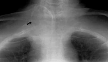
Coned apical radiograph of the upper thorax shows curvilinear calcification in a thyroid adenoma, at the root of the neck, on the right side.
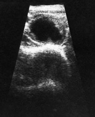
Sonogram demonstrates a benign cystic lesion in the thyroid, with a surrounding halo and ragged walls.
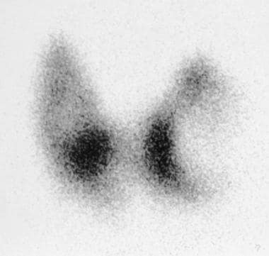
Technetium-99m pertechnetate thyroid scan shows a large cold nodule in the left lobe of the thyroid and a further, smaller cold nodule in right lobe.
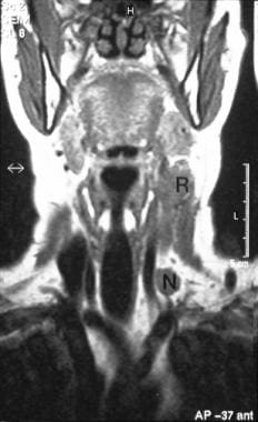
A 56-year-old man underwent subtotal thyroidectomy for a familial medullary carcinoma 2 years previously. On routine follow-up examination, a mass was felt in the thyroid. Coronal, T1-weighted magnetic resonance imaging scan shows a carcinoma recurrence (R) and lymph node (L) metastases.