
Practice Essentials
The brachial plexus represents a complex network of nerves formed from the ventral rami of the lower cervical nerves (C5-C8) and the greater portion of the ventral ramus of the first thoracic nerve (T1). The ventral rami or roots coalesce to form trunks, divisions, cords, and branches, which provide motor and sensory innervation to the upper limb and thorax.
Pathology of the brachial plexus is widely varied, but it tends to be due to similar mechanisms based on age. In neonates, the most common pathology of to the brachial plexus is related to birth trauma. In adolescents and young adults, pathology is often due to high-speed trauma, such as motor vehicle accidents. In adults, neoplasm (intrinsic or extrinsic masses) or postradiation injury frequently is the cause. Traumatic injuries to the brachial plexus are associated with weakness and paresthesias of the upper extremity on the affected side. Thorough neurologic examination can be performed to localize the injury and to help pinpoint the location of pathology. (See the image below.)
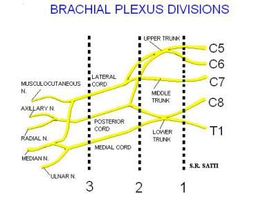
Diagram of the brachial plexus.
Clinically, brachial plexopathy commonly mimics the symptoms and signs of cervical spondylosis-related radiculopathy, and it is important to differentiate these pathologies, as the management strategies can differ. The deep location of the brachial plexus and its complex architecture make it difficult to diagnose, characterize, and treat brachial plexus lesions, often leading to inconclusive electrodiagnostic (ED) testing.
Indications for brachial plexus MRI include symptomology, pain, neural deficit, or muscular atrophy that may be due to brachial plexus pathology, postradiation treatment evaluation, or preoperative evaluation of known intrinsic or extrinsic lesions within the neck, clavicular region, or axilla.