
Cyanosis and the Clinical Assessment of Hypoxemia
Cyanosis is a bluish or purplish tinge to the skin and mucous membranes. See the images below.
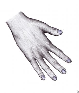
Cyanosis of nail beds.

Cyanotic lips in a woman with hypoxia.
Before the era of rapid blood gas analysis, clinicians often assessed hypoxemia on clinical grounds alone, primarily by looking for cyanosis in the perioral area and fingers.
Clinical assessment of hypoxemia is now known to be notoriously unreliable.
A host of factors, from natural skin pigment to room lighting, can affect detection of cyanosis. As with many other physical examination findings, significant interobserver variation occurs in detecting cyanosis.
Physicians may diagnose cyanosis as an indicator of hypoxemia when the patient has normal oxygen saturation; alternatively, physicians may miss cyanosis when it should be present (the patient has very low oxygen saturation with normal hemoglobin).
Approximately 5 g/dL of unoxygenated hemoglobin in the capillaries generates the dark blue color appreciated clinically as cyanosis. For this reason, patients who are anemic may be hypoxemic without showing any cyanosis.
Ancillary signs and symptoms of hypoxemia (eg, tachycardia, tachypnea, mental status changes) are nonspecific and of no value in reliably detecting hypoxemia. For example, patients may be dyspneic at rest for reasons other than hypoxemia (ie, they have normal PaO2 and SaO2). Conversely, many patients who are chronically hypoxemic (low PaO2 and/or low SaO2) are perfectly lucid and without any obvious physical signs of their low oxygen state (at least while at rest).