
Overview
The paranasal sinuses are air-filled spaces located within the bones of the skull and facial bones. They are centered on the nasal cavity and have various functions, including lightening the weight of the head, humidifying and heating inhaled air, increasing the resonance of speech, and serving as a crumple zone to protect vital structures in the event of facial trauma.
Four sets of paired sinuses are recognized: maxillary, frontal, sphenoid, and ethmoid (see the image below).
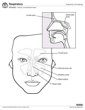
Sinuses, anterior and sectioned views.
Maxillary sinus
The maxillary sinus is the largest paranasal sinus and lies inferior to the eyes in the maxillary bone. It is the first sinus to develop and is filled with fluid at birth. It grows according to a biphasic pattern, in which the first phase occurs during years 0-3 and the second during years 6-12. The earliest phase of pneumatization is directed horizontally and posteriorly, whereas the later phase proceeds inferiorly toward the maxillary teeth.
This development places the floor of the sinus well below the floor of the nasal cavity. The shape of the sinus is a pyramid, with the base along the nasal wall and the apex pointing laterally toward the zygoma. The natural ostium of the maxillary sinus is located in the superior portion of the medial wall.
The anterior maxillary sinus wall houses the infraorbital nerve, which runs through the infraorbital canal along the roof of the sinus and sends branches to the soft tissues of the cheek. The thinnest portion of the anterior wall is above the canine tooth, called the canine fossa, which is an ideal entry site for addressing various disease processes of the maxillary sinus.
The roof of the maxillary sinus is the floor of the orbit. Behind the posteromedial wall of the maxillary sinus lies the pterygopalatine fossa, a small inverted space that houses several important neurovascular structures and communicates with several skull base foramina. The infratemporal fossa lies behind the posterolateral wall of the maxillary sinus.
The maxillary sinus is supplied by branches of the internal maxillary artery, which include the infraorbital, alveolar, greater palatine, and sphenopalatine arteries. It is innervated by branches of the second division of the trigeminal nerve, the infraorbital nerve, and the greater palatine nerves.
Frontal sinus
The frontal sinus is housed in the frontal bone superior to the eyes in the forehead. It is formed by the upward movement of anterior ethmoid cells after the age of 2. Developmentally, this is the last sinus to pneumatize. Growth increases at age 6 years and continues until the late teenage years.
The frontal sinuses are funnel-shaped structures with their ostia located in the most dependent portion of the cavities. The posterior wall of the frontal sinus, which separates the sinus from the anterior cranial fossa, is much thinner than its anterior wall.
The frontal sinus is supplied by the supraorbital and supratrochlear arteries of the ophthalmic artery. It is innervated by the supraorbital and supratrochlear nerves of the first division of the trigeminal nerve.
Sphenoid sinus
The sphenoid sinus originates in the sphenoid bone at the center of the head. It arises not from an outpouching of the nasal cavity but from the nasal embryonic lining. The sinus reaches its full size by the late teenage years. The sphenoid sinus is variably pneumatized and may extend as far as the foramen magnum in some patients.
The thickness of the walls of the sphenoid sinus is variable, with the anterosuperior wall and the roof of the sphenoid sinus (the planum sphenoidale) being the thinnest bones. The sphenoid sinus ostium is located on the anterosuperior surface of the sphenoid face, usually medial to the superior turbinate.
The sphenoid sinus is supplied by the sphenopalatine artery, except for the planum sphenoidale, which is supplied by the posterior ethmoidal artery. Innervation of the sphenoid sinus comes from branches of the first and second divisions of the trigeminal nerve.
Ethmoid sinus
The ethmoid sinuses arise in the ethmoid bone, forming several distinct air cells between the eyes. They are a collection of fluid-filled cells at birth that grow and pneumatize until the age of 12. The ethmoid cells are shaped like pyramids and are divided by thin septa. They are bordered by the middle turbinate medially and the medial orbital wall laterally. The ethmoid labyrinth may extend above the orbit, lateral and superior to the sphenoid, above the frontal sinus, and into the roof of the maxillary sinus.
The ethmoid sinuses are supplied by the anterior and posterior ethmoidal arteries from the ophthalmic artery (internal carotid system), as well as by the sphenopalatine artery from the terminal branches of the internal maxillary artery (external carotid system).