
Overview
The standard 12-lead electrocardiogram (ECG) is one of the most commonly used medical studies in the assessment of cardiovascular disease. It is the most important test for interpretation of the cardiac rhythm, detection of myocardial ischemia and infarction, conduction system abnormalities, preexcitation, long QT syndromes, atrial abnormalities, ventricular hypertrophy, pericarditis, and other conditions.
The use of standardized terminology is recommended to allow application of ECG diagnoses across many different hospital systems and countries.
See Are You Missing Subtle MI Clues on ECGs? Test Your Skills, a Critical Images slideshow, to help identify a variety of ECG abnormalities.
History of the ECG
The ECG has its roots in the seminal work of Galvani, who famously noted the contraction of frog legs when stimulated with electricity.
Although he was incorrect in attributing this phenomenon to “animal electricity,” his findings ushered in a period of research in the interaction of electricity and biological systems.
Matteucci, and later Kolliker and Muller, demonstrated that the electrical activity of a beating heart caused synchronous contractions in muscle preparations that were connected to the heart by nerve tissue.
Gabriel Lippman developed the capillary electrometer, making it possible to measure the minute currents in biological systems.
With the Lippman electrometer, Agustus Waller performed the first ECG of a human heart at St Mary’s Hospital in London in May 1887.
Willem Einthoven improved the ECG using the Lippman electrometer and then introduced the string galvanometer, which represented a great leap forward in electrocardiography.
With Einthoven’s improvements, the now familiar P, Q, R, S, and T waves were apparent.
Not only did Einthoven reveal the ECG waveforms as we know them today, but his convention for placement of the ECG leads is still in use, with leads on the right and left arms as well as the left leg, using the right leg as an electrical ground. With these leads, he demonstrated the well-known “Einthoven’s triangle” (see the image below).
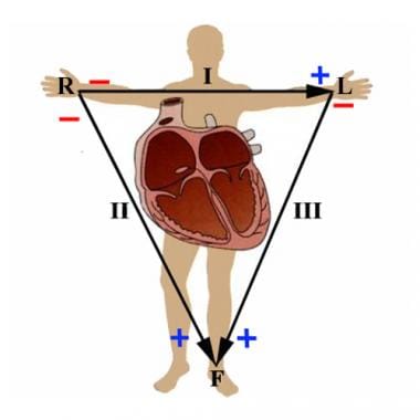
I, II and III represent the original three leads used by Einthoven. These describe the electrical vector between the right and left arms, right arm and left leg, and left arm and left leg, respectively. R, L, and F represent the augmented limb leads aVR, aVL, and aVF. They are calculated as follows: aVR = (I+II)/2 , aVL = (I-III)/2, aVF = (II+III)/2.
One of Einthoven’s students, Sir Thomas Lewis, used electrocardiography to reveal the mechanisms behind atrial fibrillation and other conduction abnormalities. In the United States, Wilson and Goldberger pioneered the use of additional leads, namely aVR, aVL, aVF, and the 6 precordial leads (see the images below).

The hexagonal reference system is the standard method of determining the axis of electrical activation of the heart. As an example, depolarization parallel with lead II would yield a value of 60º, whereas a depolarization vector in the opposite direction would be -120º.

Proper placement of the precordial leads for standard 12-lead electrocardiography.
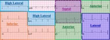
A 12-lead electrocardiogram is divided into the following four main contiguous lead groups: inferior, lateral, septal, and anterior.
After these advancements, the ECG as we know it today was created and, except for advances in the technology, it has changed little over the last 50 years.
Additional resources
For more information, see the following resources:
For a concise review of the recognition of artifacts which may be introduced by lead misplacement, see Surawicz, B, Knilans T, Chou TC. Chou’s Electrocardiography in Clinical Practice: Adult and Pediatric. 5th ed. Philadelphia, Pa: WB Saunders and Co; 2001:576-82.
For further information on the technical standards of ECG recording devices, the see the Association for the Advancement of Medical Instrumentation (AAMI) and the American National Standards Institute (ANSI).