
Overview
Tympanocentesis is a minor surgical procedure that refers to puncture of the tympanic membrane with a small gauge needle in order to aspirate fluid from the middle ear cleft or to provide a route for administration of intratympanic medications. The procedure was described in 1768 and has been used since to treat acute otitis media (AOM).
It was particularly popular in the preantibiotic era, but its use has since declined. It is now used mainly for the management of complex cases that have not responded to antibiotic therapy,
as well as facilitating the delivery of medication directly to the middle and inner ear.
Relevant Anatomy
The primary functionality of the middle ear (tympanic cavity) is that of bony conduction of sound via transference of sound waves in the air collected by the auricle to the fluid of the inner ear. The middle ear inhabits the petrous portion of the temporal bone and is filled with air secondary to communication with the nasopharynx via the auditory (eustachian) tube.
The tympanic membrane (TM) is an oval, thin, semi-transparent membrane that separates the external and middle ear (tympanic cavity). The TM is divided into 2 parts: the pars flaccida and the pars tensa. The manubrium of the malleus is firmly attached to the medial tympanic membrane; where the manubrium draws the TM medially, a concavity is formed. The apex of this concavity is called the umbo. The area of the TM superior to the umbo is termed the pars flaccida; the remainder of the TM is the pars tensa (see the image below).
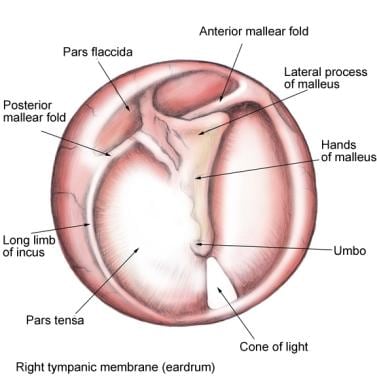
Tympanic membrane (TM): pars flaccida (superior to insertion manubrium) and pars tensa (remainder of TM).
For more information about the relevant anatomy, see Ear Anatomy.