
Background
In 1962, Headington and French first described trichilemmoma as a benign neoplasm with differentiation toward pilosebaceous follicular epithelium,
or outer root sheath. Although unusual, at times, a central zone of desmoplasia may develop and thus be termed desmoplasia trichilemmoma.
While benign in nature, the significance of trichilemmoma resides in the association with Cowden disease (ie, multiple hamartoma syndrome), nevus sebaceous, and the need to differentiate trichilemmomas from other more aggressive cutaneous tumors, such as trichilemmal carcinoma.
Clinically, trichilemmomas present as well-defined, smooth, asymptomatic papules or verrucoid growths. They may appear as a solitary or multiple lesions, and they usually are found on the head and face (see the image below). These papules often mimic a basal cell carcinoma or a verruca.
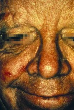
A patient with trichilemmoma papules on the face.
Trichilemmomas are often reported in association with several other neoplasms. Trichilemmoma is most commonly found secondary to nevus sebaceous of Jadassohn.
. Nevus sebaceous is classified as a congenital cutaneous hamartoma that presents on the scalp or face sometime between birth and childhood development.
Several articles have reported trichilemmomas also appearing alongside trichoblastoma, sebaceous adenoma, and syringocystadenoma papilliferum.
When multiple trichilemmomas are present, Cowden disease should be suspected, especially if associated with oral fibromas, goiter, gastrointestinal polyposis, thyroid disease, or a family history of breast cancer.